Superpower X-ray light
On the 100th anniversary of Wilhelm Conrad Röntgen's death on 10 February 2023: from his first X-ray image to a unique X-ray microscope in Hamburg
The world's first X-ray image shows the bones of the hand of Wilhelm Conrad Röntgen's wife. He took the image more than 100 years ago. Today, X-rays reveal much more than bones. Since their discovery, "X-rays" have had an impressive career: customs officials use them to screen lorries and containers; at the airport, they penetrate every piece of luggage, and X-ray fluorescence analysis is used to discover hidden layers of paint in historical paintings.
However, at DESY in Hamburg, X-rays are taken to the extreme: the X-ray light at DESY is about 1 million times brighter than a standard X-ray machine at your GP’s. What is more, the X-rays generated by the X-ray light source PETRA III are up to 5,000 times finer than a human hair. Some 6,000 researchers study extremely small samples every year at DESY. Samples range from tiny crystals of proteins for medicine to quantum materials for the hard disks of the future.

But even that is not enough for the researchers. They want to understand more clearly what happens at the smallest, molecular level. They are therefore planning a new, unique X-ray microscope: PETRA IV. More than 80 engineers and scientists have developed the concept for PETRA IV.
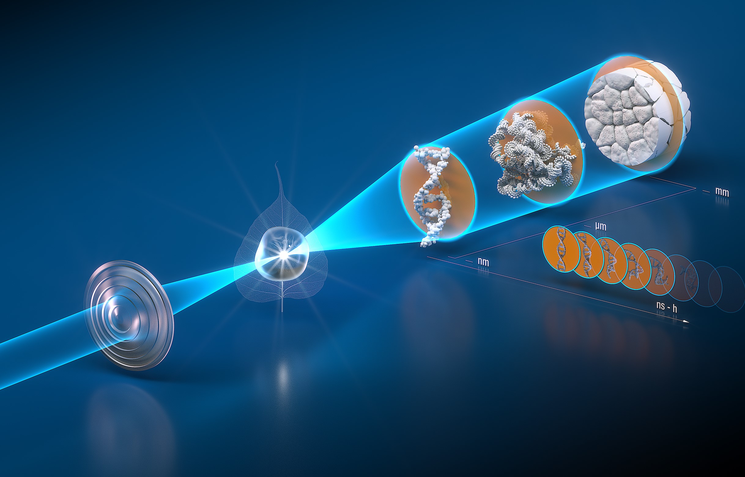
The new X-ray beam can be focused down to a nanometre in diameter. The advantage of this beam property, which has been improved by a factor of 1000 compared to PETRA III, is that the smallest processes - for example in the nucleus of a cell or in crystals of a metal - can be monitored in real time. Due to the PETRA accelerator's circomference of 2.3 kilometres, it will be possible to bundle the beam down to very short X-ray wavelengths. There is no comparable X-ray light source in the world that can achieve this brilliance. Scientists will be able to conduct experiments at PETRA IV that are only possible at this facility.

There is great demand for this facility; there are many research questions that could be answered with X-ray light.
How do bacteria manage to become immune to antibiotics? Why does a crack in the railway track propagate? Where are possible fractures in the semiconductor of the chip? Today's analytical methods do not always provide satisfactory answers to these questions. With the new light from PETRA IV, the researchers hope to clarify these questions in the future.
Would Wilhelm Conrad Röntgen have guessed what groundbreaking insights his technology would one day provide?

Heidrun Hillen
I am happy to answer your questions about PETRA IV.

